Powered By Blogger

Sunday 24 May 2015

Zombies!!:p REMARKABLE!"Judge me by my size, do you? And well you should not.”

Making zombies of ants, Swiss cheese of snails and Sherpas of sheep and cows, the lancet liver fluke proves that you don’t need to be big to be powerful.

Growing up to 10 mm by 2.5 mm (0.5 x 0.1 inches), Dicrocoelium dendriticum is a parasitic flatworm (also known as a trematode) that begins life as eggs living in the poop of (generally) cows or sheep. Shortly after being deposited, the eggs are ingested by a snail (such as Zebrina spp. or Cionella spp.) where they hatch into larva (miracidia). In this form they burrow through the snail’s gut and rest in its connective tissue where they develop into a second larval stage (sporocysts). Now they move to the digestive gland where they bear female sporocysts, that themselves produce yet another larval stage (cercariae). These last travel to the snail’s respiration chamber from which the snail finally rids itself of the parasite when it exits as a slime ball.
SMALL STORY  OF THE BASIC LIFE CYCLE OF LANCET LIVER FLUKE

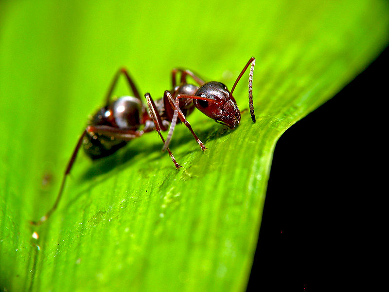
As appetizing as that sounds, it’s no wonder that an ant (such as Formica fusca) will soon wander by and eat it. Once in the ant’s intestine, the cercariae are released from the ball and most migrate to the ant’s main body cavity (hemocoel) where they transform into a fourth larval stage (metacercariae).
However, one evil genius metacercaria does not join its siblings in the ant’s hemocoel, but rather travels to a cluster of nerve cells (sub-esophageal ganglion), where it “takes control of the ant’s actions by manipulating these nerves.” When night falls, the metacercaria then directs the ant to climb to the top of a blade of grass, where it stays until the morning; the ant will repeat this nightly ritual (at the behest of its puppet master) until it is eaten by a grazing animal.

The fluke has now reached the body where it will finally mature (called the primary or definitive host). It makes its way from the animal’s small intestine to its bile duct, where it reproduces and makes eggs, the latter of which are pooped by the animal into the field to start the cycle all over again.
D. dendriticum is found throughout the world and although it primarily infects sheep, cows, snails and ants, it has been known to inhabit pigs, goats, alpacas and llamas. In fact, while rare, human infections are not unknown, with the flukes infesting human bile ducts. Usually those infected suffer from only mild symptoms include bloating and diarrhea, although some suffer from enlargement of the lining of the bile ducts (biliary epithelium) that, along with a growth of fibrous tissue, can cause the liver to swell (hepatomegaly) and cirrhosis. Note that human infection is extremely rare, as you might imagine given the way one would have to acquire the parasite.  For instance, one documented case happened only after a man drank water that had infected ants in it.
Regardless, you have to hand it to the lancet liver fluke that, during the course of its life, commonly forces three different animals to do its bidding, and completely bends the ant to its will. As another tiny powerhouse once said, “Judge me by my size, do you? And well you should not.”

LEAD is bad for humans!


Given that humans have been using lead in various product for over 8,000 years (with the first known mining of it in Anatolia around 6500 BCE), you might be surprised to learn that we have known that lead is dangerous and shouldn’t be trifled with since at least 150 BC, when its effects on the human body were noted by famed Greek physician Nicander of Colophon. Nicander even went so far as to describe the metal as “deadly”, writing extensively on the crippling effects it has on the human body in his work, Alexipharmaca.
Further, Greek physician Pedanius Dioscorides noted in the first century AD: “Lead makes the mind give way”.
To quote the Occupation Safety and Health Research Institute: “Lead poisoning is one of the earliest identified and most known occupational disease. Its acute effects have been recognized from antiquity.”

So what exactly does lead poisoning do to the body? Well, depending on how much of the substance gets into your body (and it doesn’t take much, particularly for children), it can cause everything from constipation to permanent reduction in your IQ and mental capacity. It also can potentially fundamentally change a given person’s personality, causing them to be irritable and suffer from erratic mood-swings and fatigue without warning; cause a reduction in sperm count and infertility; stunted growth (in children); miscarriages; and a whole slew of other terrifying symptoms.
So that’s what it can do, but how? Why is lead so dangerous to the human body while we can safely ingest many other types of metals, like iron, without worry (and, in fact, need some of them to survive)? While research is ongoing into the full effects and mechanisms involved in lead poisoning, what we do know is that a lot of the damage is due to the fact that important things like zinc, calcium, and iron in the body can ultimately get replaced by lead in many key biochemical reactions, if lead is present.  Unlike these other metals, though, while lead is happy to bind and interact with various critical enzymes, the result isn’t the normal reaction you need.
For example, with calcium, as noted in this paper on the Mechanisms of Lead Neurotoxicity, lead has a nasty habit of being able to mimic, or in some cases straight up inhibit the actions of calcium in natural biological reactions that take place within the human body, inhibiting neurological function, among other things.
Lead also can damage DNA, as well as your cell membranes, the latter of which, combined with the fact that it also interferes with heme synthesis, can result in anemia among a host of other problems. It can interfere with the ability for your body to synthesize vitamin D, which comes with yet another host of its own problems if you don’t have enough. It also causes a few different problems with your immune system; interferes with metabolism of bones and teeth; can cause abnormal calcium build up within cells… the list goes on and on and on.

If that wasn’t bad enough, lead can easily find its way into almost any part of your body once introduced, whether by breathing it in, ingesting it, or (very rarely) via skin absorption.
From this, you might find it completely unsurprising that, unlike many other poisons, according to the Centers for Disease Control and Prevention lead is so toxic to humans that, “No safe blood lead level has been identified.”
So, in short, lead is bad for you because, though lead has no useful function in your body, it’s happy to jump on in and give it its best college try, interacting up a storm with various enzymes, failing the whole way at producing the reactions that are needed for normal body function. But what it lacks in end result, it makes up for in staying power. You see, the half-life of lead in the body is quite long- weeks in your blood, months in your soft tissues, and years in your bones; and by years we mean up to two to three decades. Who needs proper enzymatic function anyway?..................:p...

Saturday 23 May 2015

BLACK BOARD CHALKS NOT CHALKS!


Ubiquitous in many classrooms since the 19th century, chalk and chalkboards are familiar to most of us. White, powdery and prone to sticking to those surfaces where it is put (and just as easy to wipe away), chalk and its accompanying board are excellent instructional aids. Notably, however, most chalk today isn’t technically chalk at all, but gypsum.
Chalk and gypsum have both been mined since ancient times. Chalk (calcium carbonate) has been found in cave paintings that date back to 40,000 BC, while gypsum (calcium sulfate) has been used as a mortar for construction since the dawn of civilization, and is even found in the Egyptian pyramids.

Similar and yet distinct, chalk is a base (an alkali that neutralizes acids) that is composed of calcium and oxygen combined with carbon (CaCO3), while gypsum is a salt (the product of a base and acid reacting and both becoming neutralized), made up of calcium and oxygen combined with sulfur.
Both are believed to be formed in similar fashion. Chalk is a limestone deposit created as plankton (tiny marine organisms) concentrate calcium in their bodies while living, then leach the calcium out after they die and settle onto ocean floors; over millennia, large deposits are formed, and as the seas recede, chalks deposits remain.
Gypsum’s origins are similar, but in addition to being comprised of the calcium produced by the deaths of millions of plankton, gypsum also contains some of the salt that was left behind as the ocean evaporated.
Traditionally chalk has been used for drawing and writing, and by the end of the 18th century, with advances in slate quarrying (slate was originally used for writing tablets and blackboards), writing slates covered with chalk letters, symbols, numbers and figures were commonplace. Gypsum on the other hand, had been used primarily in construction, such as for the aforementioned mortar, as well as in the manufacture of windows.
Nonetheless, both are susceptible to a process that produces sticks of themselves that, when pressed against certain services, leave washable marks.
After quarrying, each is crushed, ground, washed and sifted. With gypsum, it must also be dehydrated in a process that involves high temperatures to reduce its water content from nearly 21% to about 5-6%; to make classroom chalk, the material is mixed, again, with water (and colored pigments, if desired), and to produce more exotic pastels, such as used for art drawings, pigments as well as clays or oils are also added. For the former, the chalk is baked, while with the latter, it is air-dried.

It’s not clear why gypsum has replaced chalk for writing on blackboards (which today are mostly green, but that’s another story). While historically chalk has been remarkably dusty, modern manufacturing methods, including baking the chalk and coating it with products like shellac, have reduced the problem for both materials.
The most likely reason is that gypsum is abundant, easily mined and processed in enormous quantities. The mineral is mined in more than 90 countries, including Canada, Mexico, Spain and the United States. In the U.S., 19 states (notably California, Iowa, Nevada, Oklahoma and Texas) have surface-mining operations, and the U.S. alone produces more than 30 million tons of the stuff each year.
Sometimes called “the rock nobody knows,” gypsum can easily be turned into a fine powder (by dehydrating it), while it also has the remarkable quality of being “the only natural substance that can be restored to its original rock-like state by the addition of water alone.”
Since it can be reconstituted, it can be fashioned into any of a myriad of shapes and modified for various uses. Common applications include plaster of Paris, creating clay molds from which a variety of plastic products are formed (such as plastic cups and plates), the manufacture of glass, as an ingredient in cement and in drywall (this last has been a boon to humanity as gypsum is naturally fire resistant).
Non-toxic, gypsum also pops up in fertilizers and soil conditioners, hair products, to make plasters of teeth, to grow mushrooms, brew beer and bind tofu (which also has the added benefit of making bean curd an excellent calcium source).

Thursday 21 May 2015

The origin of DNA testing.DNA finger printing helped to solve mystry cases???

DNA testing...is the most important study in today's world. So how did this DNA finger printing came into existence. Lets see how these finger printing technology helped to solve cases.

Eureka Moment
On September 10, 1984, geneticist Alec Jeffreys, 34, was working in his lab at the University of Leicester, in central England. More precisely, he was in the lab’s darkroom, studying an X-ray that had been soaking in a developing tank over the weekend. The X-ray was the result of a process through which recently discovered DNA sequence anomalies appeared on a sheet of film as rows of black lines interspersed with blank spaces- almost like bar codes. The particular X-ray he was looking at showed DNA “bar codes” from three people: one of his technicians and her mother and father.
Jeffreys had no idea what to expect from the X-ray- he was just inventing the process, hoping to see evidence of change to specific regions of DNA between the parents and their daughter.  But after looking at the blurry mess of dark and light spaces for a few moments, he suddenly realized that, completely by accident, he had discovered a way to tell if people were related. “It was an absolute Eureka moment,” he told a reporter in a 2009 interview with The Guardian newspaper. “It was a blinding flash. In five golden minutes, my research career went whizzing off in a completely new direction.”
After the Eureka
What Jeffreys saw in that blurry X-ray: 1) each of the three family members had their own unique “bar code,” 2) all three of the family members’ bar codes related to one another (which makes perfect sense, as each of us gets our DNA as a combination of our parents’ DNA), and 3) the relationships were plainly visible. Jeffreys quickly realized that his findings would have implications regarding paternity.  With such technology you could prove with scientific certainty whether someone was- or wasn’t- someone else’s child. Or even whether they were closely related. The technology could also be of use in criminal cases where the perpetrators left blood or other biological evidence behind.
Jeffreys had apparently discovered something extraordinary- but what to do with it? Surely it would take decades for it to have any applications in the real world, he thought. So he simply kept working on what he dubbed his “DNA fingerprint” process, trying to improve it. Meanwhile, he wrote a scientific paper titled “Individual-Specific Fingerprints of Human DNA,” which was published in the scientific journal Nature in July 1985.
Two weeks later, he got a phone call.

Test Case: Paternity test.
The call came from a London lawyer who told Jeffreys she’d read a newspaper article about his “DNA fingerprinting” and wondered if it could be used in an immigration case she was handling. A British-Ghanaian woman’s 13-year-old son had gone to stay with her estranged husband in Ghana for some time, and when he returned, British authorities didn’t believe it was him. They thought the family was trying to sneak someone else- possibly a cousin- into the country on the son’s passport, and they wanted to deport the boy. Could Jeffreys prove that the child was the woman’s son?

Jeffreys agreed to give it a try. He took blood samples from the mother, three of her other children, and the boy in question, and made DNA bar codes for each of them. His conclusion: The boy was definitely the woman’s son. The lawyer presented the evidence to the British Home Office, and even though DNA testing had never been used in a case before, they were convinced. The boy was legally accepted as the woman’s son and allowed to stay in the country. Not only that, British immigration officials said they would allow DNA testing to decide any future cases that had paternity questions. The British Home Office had, perhaps without realizing it, made the brand-new, still not widely understood use of DNA testing a legally legitimate procedure.
 AFTERMATH:
News of these events made global headlines. Within a year, DNA fingerprinting- now known as DNA profiling- was being used in the United States, and in just a few more years it was considered a standard part of forensics almost everywhere in the world. And not just to find out whodunnit- but also to determine who-didn’t-dunnit.
Jeffreys is still a professor at the University of Leicester, although he is now known as Sir Alec Jeffreys. He was knighted by Queen Elizabeth II in 1994 for “Services to Science and Technology.” He has received numerous other awards for what turned out to be one of the most momentous scientific discoveries of modern times. And it brought him some well-deserved fame: “Literally every two or three days I get an e-mail,” he said in 2009, “mainly from the States, from school kids saying, ‘I’ve got to do a project on a famous scientist, so I’ve chosen you,’ and I love that. I always respond.”

SOME INTERESTING FACTS:
  • It May seem elementary to CSI fans, but after his discovery on that fateful Monday morning in 1984, Jeffreys had no idea if the DNA in a bloodstain would be usable in his process. So he did the only thing a good scientist could: “I spent the next two days cutting myself and leaving blood marks around the laboratory. Then we tested those bloodstains.” (It worked, of course.)
  • Jeffreys’s original X-rays- the ones mentioned at the start of the story, with the bar codes of the three family members- actually held 11 such codes. The other eight were made from the DNA of animals, including a mouse, a cow, and baboon. And in case you were wondering, DNA testing works the same for animals as it does for humans.

Wednesday 20 May 2015

Microbesss!

 The nature of symbiotic associations in our human body

Normal microbiota of Conjuctiva:
1.Staphylococcus
2. Haemophilus species
3.Staphylococcus aureus
4.Streptococci
Normal microbiota of outer ear:
1.Staphylococcus
2.Diptheroids
3.Pseudomonas species
4.Enterobacteiaceae
Normal microbiota of Stomach:
1.Streptococcus
2.Staphylococcus
3.Lactobacillus
4.Peptostreptococcus
Normal microbiota of Skin :
1.Coagulase-negative streptococci
2.Diptheroids(including propionibacterium aces)
3.Staphylococcus aureus
4.Streptococci
5.Bacillus
6.Malasseria furfur
7.Candida species
8.Myobacterium species
Normal microbiota of Urethra:
1.Coagulase-negative streptococci
2.Diptheroids
3.Streptococci
4.Mycobacterium species
5.Bacteroides species and fuso bacterium
6.Peptostreptococcus species
Normal microbiota of Nose:
1.Lactobacillus 
2.Viridans streptococci
3.Staphylococcus aureus
4.Neisserio species
5. Haemophilus species
6.Streptococcus pneumoniae
Normal microbiota of  small intestine:
1.Lactobacillus sp.
2.Clostridium sp.
3.Bacteriods sp.
4.Mycobacterium sp.
5.Enterobacteriacae
Normal microbiota of mouth and oropharynx:
1.Viridians streptococci
2.Veinella sp.
3.Fusobacterium sp.
4.Treponema sp.
5.Porphyromonas sp and prerotella sp.
6.Neisseria sp and Branhamella catarrhalls
7.Streptococcus pneumoniae
8.Beta-hemolytic streptococcus.
9.Candida sp.
10.Haemophilus sp.
11.Diptheroids
12.Actinomyces sp
13.Eikenella corrodens
14.Staphylococcus aureus
Normal microbiota of large intestine:
1. Bacteriods sp.
2.Fusobacterium sp.
3. Clostridium sp.
4.Peptostreptococcus
5.E.coli
6.Klebsiella sp.
7.Lactobacillus
8.Enterococci
9.Streptococci
10.Pseudomonas sp.
11.Acinetobactor sp.
12.Staphylococcus aureus
13.Mycobacterium sp
14.Actinomyces sp

Saturday 9 May 2015

Confectioner's gaze~~~~~~~:p GEMS.....MOUTH WATERING!!! BUT.......


A COMMON COATING ON CANDIES AND PILLS------- IS MADE FROM THE BODILY EXCRETIONS OF AN ASIAN BEETLE

Confectioner's gaze, also called pharmaceutical glaze,resinous gaze, pure food glaze and natural gaze, is a common ingredient in candies and pills.
By any name, its the same ingredient as SHELLAC, the chemical that they sell in hardware stores and that is used for sealing and varnishing wood floors!!!!OMG!!!!!!DANGER!!...and we are eating this!!!


SHELLAC is actually a chemical secreted by female lac bugs(Laccifer lacca) , a type of "scale insect", in order to form sheltering tunnels as they travel along the outside of trees. it is extracted for industrial use by scraping bark, bugs and tunnels off of trees in Asian forests and into the canvas tubes. The tubes are then heated to over a flame until the shellac melts and seeps out of the canvas, after which it is dried into flakes for sale.Before use in food or as varnish, the shellac must be re-dissolved in denatured alcohol.

Instead of shellac ,some food producers even use acorn protein called zein. but usage of plant product like zein is not much harmful..but what about SHELLAC......do please kindly read the ingredients before you eat something.

                beautiful.......but????????                           
laccifer lacca....
...............................................................

Wednesday 6 May 2015

The $!LVER hero


The silver hero!!!


SILVER...instantly remains us of  Ornaments , jewels, and a lots more . But what if this  metal  acts as a villan to microbes  and a hero to us. lets see the  reason behind this wonderful hero.....
Thousands of years before the discovery of microbes or the invention of antibiotics, silver was used to protect wounds from infection and to preserve food and water. The alluring metal which was fashioned into a multitude of curative coins, sutures, foils, cups and solutions all but vanished from medical use once physicians began using anti-bacterial drug agents to fight sickness in the 1940s.But now, as bacteria grow increasingly resistant to these medications and new pathogens invade hospitals, some doctors are turning once again to the lustrous element that Hippocrates prescribed for patients in ancient Greece.
The silver contains a strong antimicrobial  properties which uses many chemical processes to stop  bacteria from forming bonds, slow their metabolic rate and disrupt homeostasis. This makes the bacteria more susceptible to the power of antibiotics making it weaker. A powerful germicidal, silver is an exceptional metal in that it is non-toxic to the human body, but lethal to over 650 disease causing bacteria, viruses, fungi, parasites, and molds; while conventional pharmaceutical antibiotics are typically effective against only 6 or 7 types of bacteria. Some new strains of bacteria classified as MDR (Multiple Drug Resistant) have proven to be resistant to all pharmaceutical antibiotics, but not to colloidal silver due to different germicidal mechanisms of deactivation.  

How does the hero attack the microbes??
The silver in the form of dissolved ions makes the cell membrane more permeable and interferes with the cell’s metabolism leading to the overproduction of reactive, and often toxic, oxygen compounds. Both the mechanisms could potentially be harnessed to make today’s antibiotics more effective against resistant bacteria. The three mechanisms of deactivation that silver utilizes to incapacitate disease causing organisms are Catalytic Oxidation, Reaction with Cell Membranes, and Binding with the DNA of disease organisms to prevent unwinding. 
1.Catalytic Oxidation: Silver catalyses the oxidation reaction by absorbing oxygen on its surface and readily react with the sulfhydryl(-S-H) groups surrounding the surface of the bacteria and convert them to (R-S-S-R Bond) by removing the hydrogen .This makes the bacteria to expire by blocking the respiration.it can also react with any negative charge presented by the organisms transport or membrane proteins and deactivate them. Researchers have found that the silver compound made the bacteria  to produce more reactive oxygen species(ROS), chemicals that all  living cells make as a byproduct of metabolism. ROS plays an important role in helping cells carry out normal function. But in excess, they can damage proteins and DNA and also the cell membrane to become leaky.
the researchers were also surprised to find that the silver- antibiotic combo was able to re-sensitize bacteria that had developed a resistance to drugs. Even in vanomycin(commonaly  used antibiotic).
2.Reaction with Bacterial Cell Membranes: the silver ions reacts with the surface radicals of  imparing the respiration and blocking its energy transfer system. Specific enzymes are required for for a given biochemical activity to take place. These usually require a specific metallic atom for its function. A metal of higher valance can replace a metal of lower valance in the enzyme complex, preventing the enzyme from functioning normally. Silver, with a valance of plus 2, can replace many metals with a lower, or equal valance that exhibit weaker atomic bonding properties.
3.Binding with DNA: 12% of silver is taken up by the organism's DNA. While it remains unclear exactly how the silver binds to the DNA without destroying the hydrogen bonds holding the lattice together, it nevertheless prevents the DNA from unwinding, an essential step for cellular replication to occur .
How does it help the antibiotics??
Many of the antibiotics are thought to kill the target by producing reactive oxygen compounds but when it is boosted up with a small amount of silver these drugs could kill between 10 to 1000 times more bacteria.. the increased membrane permeability also allows more antibiotics to enter the bacterial cells, which may overwhelm the resistance mechanisms that rely on shutting the drug back out.
What does it do?
The silver ions  interfere with the cellular processes in bacteria , including disulfide bond formation , iron homeostasis and metabolism. These changes makes the cell wall more permeable, but also lead to increased  production of reactive oxygen species which can induce cell death via the dna damage.
Fights with??
Addition effect of this silver is that the antibiotics are only effective in killing the gram positive bacteria like staph and strep but when aided by silver it also killed the gram negative bacteria like food poisoning microbes and infectious microbes.Researchers have also found that silver helped to deal with two kinds of treatmentthat required repeated trips to clinical antibiotic treatment Caused by bioflims, slimy layers of microbes that coat catheters and prosthetic joints.Bacteria that lie dormant during antibiotic treatment and then resurge when it finishes, causing recurrent infection.
So silver can play a really valuable role as an adjunct to current antibiotics.Because it is proving very difficult for drug companies to find new drugs to tackle the rising problem of antibiotic-resistant bacteria,Silver has long been used to prevent and treat infections.
Can it have an Adverse effect??
 It does but when it is consumed excessively.Argyria, an irreversible condition in which the skin turns blue or gray due tothe buildup of silver particles as a result of consuming silver over long periods of time. Though the quatity is very small it leads to serious infections in individuals. Silver can be toxic to nerve cells in the brain and spinal cord, but is normally prevented from entering those areas by the blood-brain barrier. Silver has not demonstrated any evidence of carcinogenic activity. The body eliminates excess silver via the metallothiones. These ubiquitous proteins, first characterized in 1957, have the property of binding with heavy metals, such as silver, into metal-thiolate-cluster structures which aid in transportation, storage, and elimination of nonessential trace metals which enter the body. 
Silver has been used as an antibiotic  since the days of  Hippocrates. Antibiotic resistance is running rampant these days. More and more bacteria strains are evolving to beat our treatments, allowing infections to persist and wreak havoc on our health. The trickiest types of infections are those that involve Gram-negative bacteria. These strains, which include E. coli and Salmonella, have an almost impermeable cell wall that protects them from antibiotics. This is why illnesses like meningitis and urinary tract infections can be tough to get over. Many findings have revealed that silver stages a multi-targeted attack on even the best-armed bacteria.
 This is a very exciting discovery for the medical world with the possible uses and applications for this precious metal continuing to grow.